
Powered By



If you have dry age-related macular degeneration (AMD), your eye doctor may have found drusen during an eye exam before you noticed any changes to your central vision. Drusen are a common finding in dry AMD.
Drusen are deposits of material that build up under the retina. Not all drusen are the same, and different types can be linked to different risks of AMD getting worse.
In this article, we’ll break down key differences between hard and soft drusen. We’ll also cover several drusen subtypes and explain the tests that help eye doctors detect and monitor them.
Drusen are deposits that contain fats and proteins and collect under the retina. On a dilated eye exam, they appear as yellow deposits and can be roughly the size of the head of a pin.
Drusen often don’t cause symptoms on their own. However, large drusen or drusen near the center of the retina may distort vision. Your eye doctor might find them during a routine eye exam before you notice symptoms of AMD.
Your risk of AMD getting worse generally increases when drusen are larger or more numerous. Drusen are thought to be part of changes that damage the retina and its supporting tissues over time, which can lead to central vision loss.
You can’t see drusen yourself, so regular eye exams are important for monitoring AMD. Your doctor may also recommend checking your vision at home with an Amsler grid.
 What Are the Key Differences of Hard vs. Soft Drusen?
 Hard drusen and soft drusen are the two main types of drusen. Their different features can help you and your eye doctor determine which type you have and estimate your risk of AMD progression and vision loss.
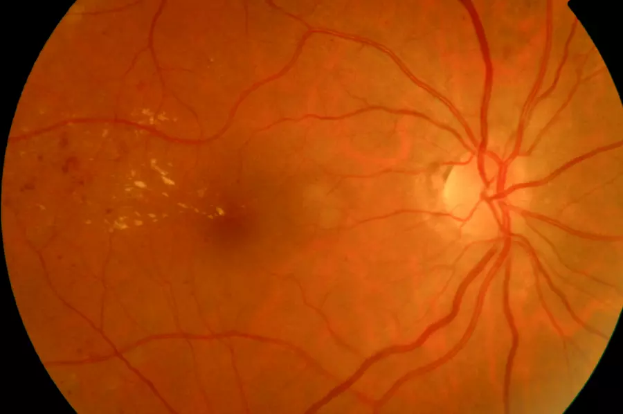
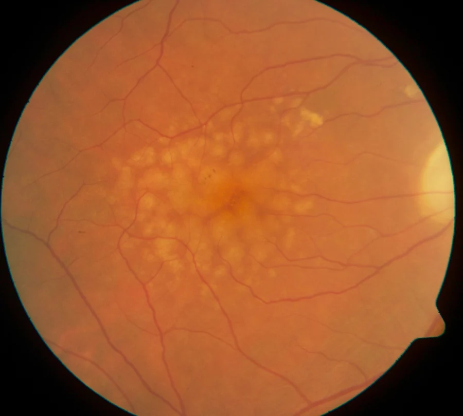
Hard drusen, which are common and usually age-related, are smaller than soft drusen. Hard drusen are also sometimes called “small drusen” because they’re less than 63 microns in diameter. Soft drusen are larger and generally more than 125 microns in diameter.
Hard drusen tend to be round with well-defined borders. On a dilated eye exam, their edges are usually easy to see.
Soft drusen on the other hand, have borders that aren’t as well defined. They’re usually moundlike and raised, and they may cluster together.
Hard drusen are common with aging, and having a few small hard drusen doesn’t necessarily mean you have AMD or will develop it later. If you only have a few hard drusen, your risk of central vision loss over the next five years is slim.
Small hard drusen can grow or develop features linked to a higher risk of AMD over time. For that reason, your eye doctor may want to monitor them even if they’re not causing symptoms.
Soft drusen, especially when they’re large or numerous, are much more likely to affect vision and cause AMD progression. The exact risk depends on several features in both eyes, including the size and number of drusen and whether there are changes in the retinal pigment. People with intermediate AMD may also have an increased risk of developing advanced AMD, including wet AMD or geographic atrophy.
Soft drusen and hard drusen aren’t the only categories used to describe drusen in dry AMD. Other subtypes can give eye doctors more information about your AMD and risk of progression.
Cuticular drusen are also called basal laminar drusen. They’re very small and tend to appear in clusters.
Cuticular drusen are typically from 25 to 75 microns in diameter, but their size can change over time. Some may shrink, whereas others may grow and fuse together. Having many cuticular drusen may raise your risk of AMD progression.
Reticular drusen are arranged in a network or lacelike pattern and are usually easier to see under blue light. Unlike typical drusen, which form below the retinal pigment epithelium (RPE), reticular drusen form above the RPE. They increase the risk of advanced AMD in both the eye where they’re found and the other eye.
Calcified drusen contain calcium-rich minerals and can look glistening or glittery during an eye exam. Among these three subtypes, calcified drusen were the least common in a large study and became more common with age. Like cuticular and reticular drusen, they’re associated with more advanced AMD stages.
You can’t tell that you have drusen just by looking at your eye, but an ophthalmologist can detect them using specialized tools. Drusen are often first discovered during routine eye exams.
It’s important to stay on track with regular eye exams so your eye doctor can detect drusen and monitor them over time. Eye doctors may use a dilated eye exam and imaging tests to look for drusen and other signs of AMD.
An optical coherence tomography (OCT) scan can show cross-sections of the retina and underlying drusen. This eye imaging test gives your eye doctor detailed pictures of the back part of your eye.
An OCT scan directs infrared light reflected from the back of your eye to create these cross-section images. Your eye doctor may use this noninvasive test to help diagnose and monitor AMD. Comparing OCT images over time can help your doctor look for changes in the retina and drusen.
Fundus photography is an imaging technique that produces a picture of the inside surface at the back of your eye. It gives your eye doctor an up-close look at your retina, macula, and optic nerve.
Fundus photography is often part of routine eye exams. Photos taken over time can also help your doctor look for drusen and watch for changes in your retina.
An Amsler grid test is a tool you can use at home to monitor your central vision for changes that may be a sign of wet AMD. Your ophthalmologist may recommend using an Amsler grid regularly after AMD is detected.
You should use the Amsler grid to check one eye at a time. If lines suddenly look wavy, blurry, dark, or missing, contact your eye doctor promptly because these changes may be a sign of wet AMD. Early treatment of wet AMD is linked to better outcomes.

Ask your eye doctor how often you should have eye exams to monitor drusen and AMD. Your eye doctor can tell you about the type and size of drusen in your eyes and what these findings may mean for your risk of AMD progression.
Your ophthalmologist may recommend Age-Related Eye Disease Study 2 (AREDS 2) supplements if you have intermediate dry AMD or advanced AMD in one eye. AREDS 2 supplements don’t make drusen go away, and their benefits are modest, so talk with your eye doctor about whether they’re appropriate for you.
On myAMDteam, people share their experiences with age-related macular degeneration, get advice, and find support from others who understand.
Which type of drusen have been found in your eye affected by AMD? Let others know in the comments below.
Get updates directly to your inbox.





 Continue with Facebook
Continue with Email
Continue with Facebook
Continue with Email
Continue with Facebook
Continue with Email
Continue with Facebook
Continue with Email
Become a member to get even more





 Join
Join

 Your Privacy Choices

This is a member-feature!
Sign up for free to view article comments.
We'd love to hear from you! Please share your name and email to post and read comments.
You'll also get the latest articles directly to your inbox.